

Your Complete Guide to X Ray Dental in Australia
A routine x ray dental appointment is one of the most powerful tools we have for keeping your smile healthy. Just as a doctor needs an X-ray to diagnose a broken bone, we rely on these images to see what’s happening beneath the surface of your teeth and gums—areas completely hidden from view.
Why Modern Dental X Rays Are So Important

Think of it this way: a visual check-up is like looking at the cover of a book. It gives us a good idea of the overall condition, but it doesn’t tell the whole story. Dental X-rays let us read the chapters inside, revealing the full picture of your oral health.
These detailed images allow us to see between your teeth and below the gumline. This helps us catch small issues early, long before they turn into bigger, more painful problems. It’s all about being proactive.
Seeing Beyond the Surface
At their heart, dental X-rays give us a clear map of your teeth and jaw. This lets us spot the earliest signs of trouble that might otherwise go completely unnoticed for months or even years. Without this insight, a tiny cavity can easily grow into something that requires a lot more work down the track.
Some of the key things we look for with X-rays include:
- Early Cavity Detection: We can find decay hiding between teeth or under old fillings while it’s still small and simple to fix.
- Assessing Bone Health: X-rays show us if there's any bone loss around your teeth, which is a major sign of gum disease.
- Identifying Hidden Problems: They can reveal issues like abscesses, cysts, tumours, or impacted wisdom teeth that haven't emerged yet.
- Planning for Treatment: These images are essential for accurately planning treatments like orthodontics, dental implants, and root canals.
> Think of dental X-rays as your mouth's early warning system. They provide the crucial information we need to shift from simply reacting to problems to proactively protecting your natural teeth for life.
The Role of Technology in Your Care
At Oasis Smiles Dental, we pair our clinical experience with the best tools available. That’s why we’ve brought advanced digital imaging technology into our Tamworth practice. Unlike old-school film X-rays, our modern digital sensors create instant, crystal-clear images with a fraction of the radiation.
What this means for you is a safer, quicker, and more comfortable appointment. The images pop up on our screen immediately, so we can sit with you and point out exactly what we’re seeing in real time. This helps you get a clear understanding of your own oral health and feel confident in any decisions you make about your treatment.
We use this technology not just to find problems, but to prevent them from starting. You can learn more about how we do this by exploring our commitment to technology. It’s all part of ensuring your family receives the highest standard of care for a healthy, confident smile.
The Hidden Problems Dental X-Rays Uncover
When you're in the dental chair, a visual check tells us a lot, but it’s only half the story. Think of your teeth like icebergs—most of the action is happening below the surface. That’s where dental X-rays come in, giving us a crucial look at the hidden parts of your teeth, roots, and jaw.
Without these images, small issues can easily go unnoticed. By the time you feel pain, the problem has often become much more serious, potentially leading to more complex and expensive treatments down the track.
Catching Decay Between Teeth
One of the trickiest spots for trouble is in the tight spaces between your teeth. Even the most diligent brushing and flossing can’t always clear away the plaque that builds up here, which can slowly lead to cavities. We call this ‘interproximal decay’.
An X-ray is the only way we can reliably spot this kind of decay early on. Finding it sooner means we can often fix it with a simple filling, instead of waiting until it burrows deeper and requires a root canal or even removal of the tooth.
Uncovering Deeper Infections and Abscesses
Sometimes, a deep cavity or a tiny crack in a tooth can let bacteria in, leading to an infection right at the tip of the root. This can form a pocket of pus called an abscess, which sits silently in your jawbone. You won't see it, and you might not even feel it at first.
On an X-ray, an abscess shows up as a distinct dark spot. Catching it early is so important because it allows us to treat the infection before it causes severe pain or spreads to the surrounding bone.
Assessing Bone Loss from Gum Disease
Gum disease (periodontitis) is a sneaky condition. It starts out quietly, maybe with a little bleeding when you brush, but it can seriously damage the bone that holds your teeth in place.
> X-rays provide an essential historical record of your oral health. By comparing your images over time, we can track even subtle changes in bone levels and intervene before significant damage occurs.
This allows us to manage gum disease at our practice proactively, helping you keep your teeth for life.
Identifying Impacted Teeth and Other Issues
Beyond just decay and infections, X-rays give us a complete picture for a whole range of other potential issues. They help us:
- Spot Impacted Wisdom Teeth: We can see exactly where wisdom teeth are positioned, even if they’re stuck under the gums or growing sideways, and assess if they need to be removed.
- Find Cysts or Tumours: While rare, X-rays can reveal abnormal growths in the jaw that might need further attention.
- Evaluate Jaw Development: For kids and teens, we use X-rays to watch how their adult teeth are coming in and to plan for any orthodontic work that might be needed.
It’s clear just how vital this technology has become. In fact, Australia's dental X-ray market was valued at USD 38.5 million in 2023 and is projected to more than double to USD 77.1 million by 2030. This trend highlights how dentists across the country are relying on better imaging to improve patient care, which you can read more about in this dental industry report.
A Closer Look at the Different Types of Dental X-Rays
Just like a builder has different tools for different jobs, we as dentists rely on a few different types of X-rays. Each one is designed to give us a specific view of your teeth and jaw, helping us see exactly what’s going on. Knowing the "why" behind each type can make your dental visits feel a lot more straightforward.
Most dental X-rays fall into two main groups: intraoral (where the sensor is placed inside your mouth) and extraoral (where the imaging is done from the outside).
Intraoral X-Rays: The Close-Up View
Intraoral X-rays are the most common ones you'll have during a check-up. We use a small, comfortable digital sensor inside your mouth to get a highly detailed picture of just a few teeth at a time. This precision is perfect for spotting problems that aren't visible to the naked eye.
The two workhorses of intraoral imaging are bitewing and periapical X-rays.
- Bitewing X-Rays: These are the stars of the show during your routine clean and check. You'll bite down on a small tab, which holds the sensor perfectly in place to capture the crowns of your upper and lower back teeth in one shot. Their main job is to find cavities starting between your teeth—a place we simply can't see otherwise.
- Periapical X-Rays: Think of this as a portrait of a single tooth. A periapical X-ray shows the entire tooth structure, from the chewing surface right down to the tip of the root and the bone that holds it in place. If you come in with a toothache, this is often the first image we’ll take to diagnose issues like an abscess or a deep infection at the root.
These images are vital for catching small problems before they become big ones. For example, a periapical view helps us see if a tooth infection has reached the nerve, which might mean a root canal is needed to save the tooth. You can learn more about how we manage tooth infections in our detailed guide.
To make it even clearer, here’s a quick guide to which X-ray we typically use for common concerns.
#### Common Dental X-Rays and Their Uses
| X-Ray Type | Primary Use | What It Shows Best | | :--- | :--- | :--- | | Bitewing | Routine check-ups | Cavities between teeth, early bone loss | | Periapical | Diagnosing tooth pain | The entire tooth, including the root and surrounding bone | | Panoramic (OPG) | Overall assessment | All teeth, jaws, wisdom teeth, and sinuses in one image |
This table is just a general guide, but it shows how we select the right tool to get the information we need to care for your smile.
Extraoral X-Rays: The Bigger Picture
Sometimes we need to step back and see your entire mouth and jaw structure. That's where extraoral X-rays come in. These images are taken from outside your mouth and give us the broad view needed for planning more complex treatments.
The flowchart below shows why this bigger picture is so important—it helps us find hidden problems like decay, infections, or bone loss that could cause serious issues if left untreated.
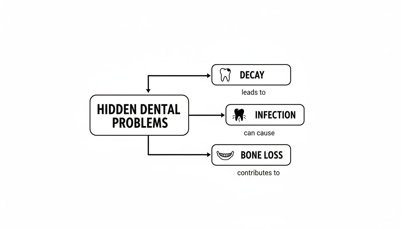
As you can see, what happens below the gumline is just as important as what you can see in the mirror.
The most common extraoral X-ray is the Panoramic X-ray, often called an OPG (Orthopantomogram). For an OPG, you’ll simply stand still for a few moments while the machine slowly rotates around your head.
> An OPG delivers a single, sweeping image of your entire mouth. It captures all of your teeth, both upper and lower jaws, your jaw joints (TMJ), and even your sinuses.
This all-in-one view is incredibly useful for:
- Assessing Wisdom Teeth: An OPG gives us a crystal-clear look at the position, development, and angle of your wisdom teeth. This helps us decide if they need to be removed and, if so, how to do it safely.
- Planning Orthodontic Treatment: Before you start with braces or clear aligners, an OPG helps us get a complete picture of your jaw and check for any unerupted, impacted, or missing teeth.
- Evaluating the Jawbone: It's our go-to for an initial look at your bone structure, which is essential when planning for dental implants or investigating sources of jaw pain.
By using the right type of dental X-ray for your specific situation, we can build a complete picture of your oral health, leading to a more accurate diagnosis and a better treatment plan for you.
Seeing Your Smile in 3D with CBCT Scans
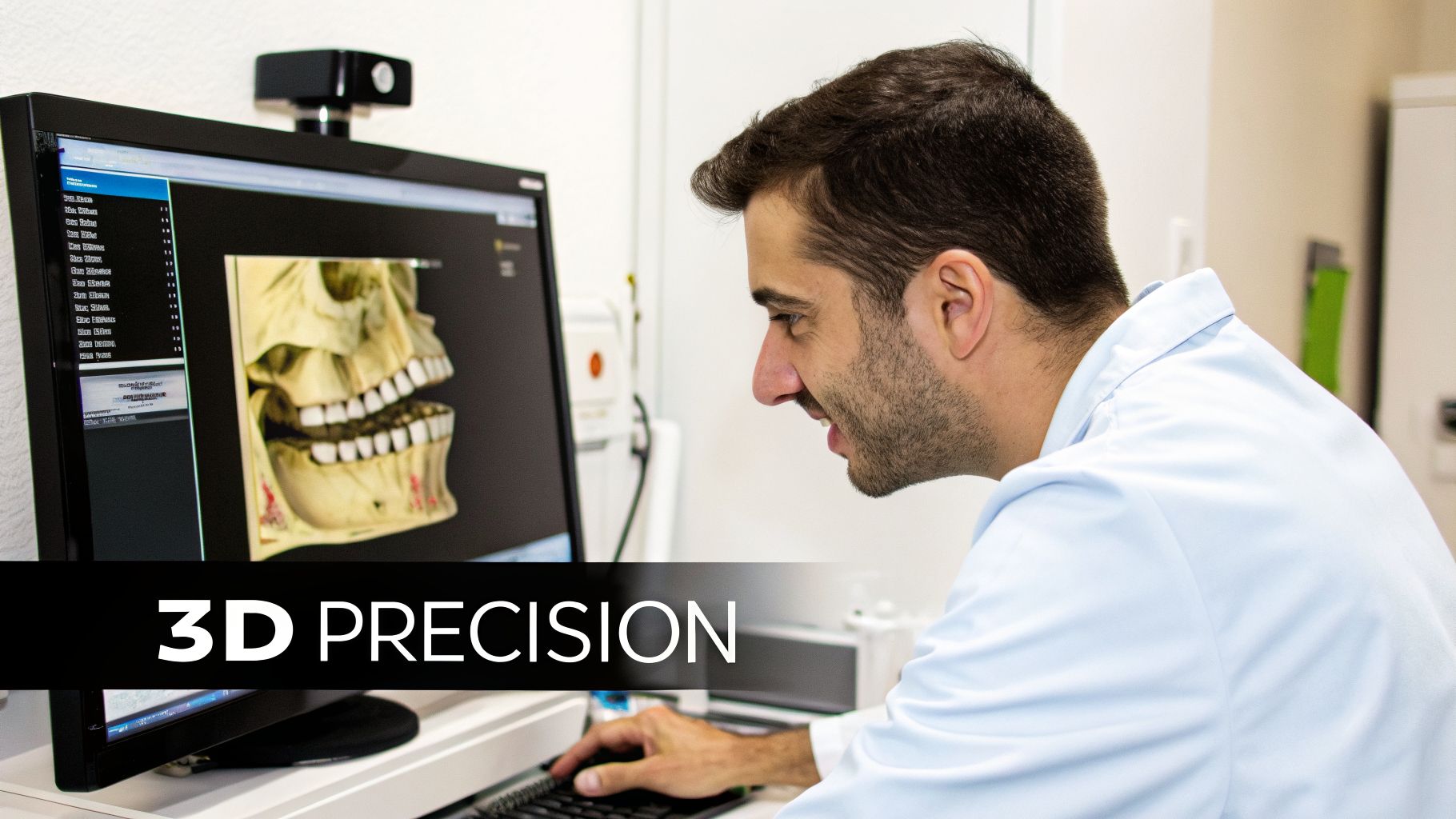
Standard X-rays give us an essential flat picture of your teeth, and for many situations, that's all we need. But sometimes, we need to see your smile from every possible angle. That’s when we turn to a much more powerful tool: Cone Beam Computed Tomography, or CBCT.
Think of a normal dental X-ray as a flat road map. It’s incredibly useful for getting from A to B, but it doesn’t show you the hills and valleys. A CBCT scan, on the other hand, is like a detailed 3D model of your teeth, jaw, and all the surrounding anatomy. Having this technology right here at Oasis Smiles Dental means we can plan complex treatments with a level of precision that just wasn't possible with 2D images alone.
How a 3D Dental Scan Changes Everything
A CBCT scanner works by rotating around your head in one quick pass, taking hundreds of pictures as it goes. Powerful software then stitches all these individual images together into a single, interactive 3D model of your mouth.
You might have heard of medical CT scans, but a dental CBCT is quite different. It uses a special cone-shaped beam that dramatically reduces your radiation exposure while still creating incredibly sharp, high-definition images. This comprehensive 3D view is an absolute game-changer for procedures that require a deep understanding of your unique anatomy.
This technology has quickly become a cornerstone of modern dentistry, and we're proud to be among the practices offering it. In Australia, the adoption of advanced x ray dental equipment is growing, with projections showing New South Wales and Victoria will account for most of the equipment revenue by 2026. This shows just how vital this technology is for providing top-tier care, and we believe our Tamworth patients deserve access to it. You can explore more about this trend in the Australian dental market if you're interested in the data.
When Do We Recommend a CBCT Scan?
We don’t need a CBCT for every patient or every check-up. We reserve it for those times when absolute precision is non-negotiable. It helps us take the guesswork out of complex situations, leading to safer procedures and much more predictable results for you.
Here are a few key situations where a 3D scan is invaluable:
- Dental Implant Planning: A CBCT scan lets us "virtually" place your implant on the computer before we even start. We can measure the exact height and width of your jawbone, pinpoint the location of nerves and sinuses to avoid them, and choose the perfect implant for a secure, long-lasting fit.
- Complex Root Canals: Some teeth have tricky, twisting, or hidden canals that a normal X-ray might miss. A 3D image illuminates the entire root system, so we can be sure we’ve thoroughly cleaned and sealed every last one, giving us the best possible chance of saving the tooth.
- Impacted Wisdom Tooth Removal: Removing a stubborn wisdom tooth, especially one nestled close to a major nerve, requires a solid game plan. A CBCT shows us the exact relationship between the tooth and the nerve, allowing us to map out the safest path for removal.
- Orthodontic Assessment: For certain orthodontic cases, a 3D view gives us a much clearer picture of how the teeth and jaws align. This helps us create a far more effective and efficient treatment plan.
> With a CBCT scan, we can measure anatomical structures with sub-millimetre accuracy. This means no surprises during your procedure and a treatment plan that is truly customised to your anatomy.
This incredible detail is particularly crucial for restorative treatments like implants. By mapping out the entire procedure digitally first, we ensure every step is optimised for safety and a successful outcome. You can learn more about how this technology supports our approach to dental implants here.
Ultimately, having this powerful diagnostic tool right here in our Tamworth practice means you get world-class treatment planning without the hassle of travelling to a separate imaging centre. It’s just one more way we work to ensure your care is as accurate, comfortable, and effective as it can be.
Of course. Here is the rewritten section, crafted to sound like it was written by an experienced and reassuring dental professional.
***
Answering Your Safety Concerns About Dental X-Rays
Let's talk about something that’s on many people's minds: are dental X-rays safe? It’s a great question, and we’re always happy to explain how we approach it. The short answer is yes—with today’s digital technology, the radiation involved in a routine x ray dental exam is incredibly low.
The easiest way to understand the dose is to compare it to the world around us. We’re all exposed to natural background radiation every single day. A standard set of dental X-rays often exposes you to less radiation than you'd get on a short flight from Sydney to Melbourne.
How We Keep You Safe During Every X-Ray
Your well-being is at the heart of everything we do. We strictly follow a principle known as ALARA, which stands for "As Low As Reasonably Achievable." In simple terms, this means we only take an X-ray when it's absolutely necessary for a proper diagnosis, and we always use the smallest dose required to get the job done right.
Every time we take an X-ray, we follow strict safety protocols:
- We shield your body. You’ll wear a heavy lead apron, and often a thyroid collar, to protect your vital organs. Think of it as a protective blanket that stops the X-ray beam from going anywhere it doesn’t need to.
- We use advanced digital sensors. Our modern digital X-ray sensors are a game-changer. They're so sensitive that they need up to 80-90% less radiation than old-school film to capture a crystal-clear image.
- We use a highly focused beam. The X-ray is a tiny, targeted beam aimed only at the specific tooth or area we need to see. This precision minimises any scatter.
To really put this into perspective, have a look at how the radiation from dental X-rays stacks up against some common, everyday sources.
Dental X-Ray Radiation Compared to Everyday Sources
This table shows just how small the radiation dose from dental X-rays is when compared to sources you encounter all the time.
| Radiation Source | Typical Dose (in microsieverts, µSv) | Comparison | | :--- | :--- | :--- | | Set of 4 Bitewing X-Rays | ~5 µSv | Less than a single day of natural background radiation | | Panoramic X-Ray (OPG) | ~10-20 µSv | Roughly equal to 1-2 days of natural background radiation | | Cross-Country Flight (e.g., Sydney to Perth) | ~40 µSv | About four times the dose of a panoramic X-ray | | Annual Natural Background Radiation | ~2,400 µSv | The average amount a person receives in a year just from the environment |
As you can see, the dose from a standard set of X-rays is just a tiny fraction of the radiation we all live with every day.
A Special Note on Children and Pregnancy
We are always extra cautious with our youngest patients and with expectant mothers.
For Children: A child’s body is still growing, so we’re especially mindful. We only take X-rays on children if they’re essential—for instance, to see decay hiding between their baby teeth, check on adult teeth coming through, or diagnose a specific problem. When we do, we use lower exposure settings tailored for their smaller size.
For Pregnant Patients: If you are pregnant or even think you might be, always let our team know. While the risk to your baby is incredibly low thanks to the lead apron and focused beam, we generally prefer to postpone any routine X-rays until after you’ve given birth, just to be on the safe side.
> The one exception is a dental emergency. If you have a serious infection or injury, the risk of leaving that problem untreated is much higher than the tiny risk from a necessary X-ray. In these urgent cases, we can safely take an X-ray with double aprons and maximum protection for you and your baby.
At the end of the day, a properly justified dental X-ray is one of the most powerful tools we have for keeping you healthy. The ability to see what’s happening beneath the surface allows us to catch problems early, prevent future pain, and plan the best possible care for your smile.
What to Expect During Your X-Ray Appointment
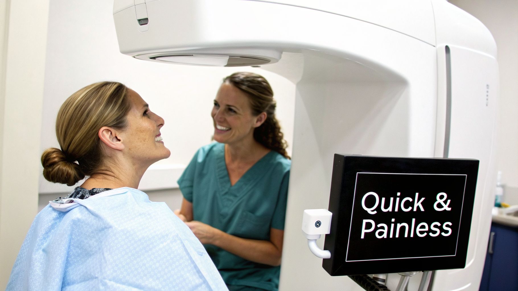
We find that knowing exactly what to expect helps take the worry out of any dental visit. A modern x ray dental appointment is a lot faster and more comfortable than you might think, and we want you to feel completely at ease from start to finish.
Our friendly dental assistant will bring you to our dedicated imaging room. First things first, we’ll get you comfortable by placing a heavy, protective lead apron over your chest and lap. This is a standard safety precaution that shields the rest of your body during the quick exposure.
Taking Close-Up X-Rays (Like Bitewings)
If your check-up calls for a detailed look, like with bitewing x-rays, the process is incredibly simple. For this, we use a small, smooth digital sensor that’s designed to fit easily inside your mouth.
- Positioning the Sensor: Our assistant will gently place the small sensor alongside your teeth.
- Biting Down: We’ll ask you to bite down softly on a little plastic guide. This just holds the sensor perfectly still so we get a crystal-clear picture.
- The "Beep": The assistant will pop out of the room for a moment to capture the image. The actual exposure is over in less than a second—you'll hear a quick "beep," and that's it. It’s completely painless, with no odd sensations.
We’ll repeat this a few times to get a complete picture of different areas, but the entire process for a full set of bitewings usually takes just a few minutes.
Getting a Panoramic (OPG) X-Ray
When we need a bigger picture of your whole jaw—perhaps to check on wisdom teeth or plan for orthodontics—we’ll use our panoramic machine. This is even easier, as nothing has to go inside your mouth.
You’ll simply stand or sit in front of the machine and rest your chin on a small, steady support. Then, the machine will slowly and quietly rotate around your head. The whole scan takes about 10-15 seconds. And just like that, you're done.
> The best part about using digital X-rays is that the images show up on our computer screen instantly. There’s no waiting around for film to develop. You and your dentist can look at them together, right then and there.
This immediate feedback lets us show you exactly what we’re looking at, whether it’s a tiny cavity starting between your teeth or the angle of a wisdom tooth. It’s a fantastic way for you to see what we see, helping you feel confident and informed about your oral health and any treatment we plan together.
Answering Your Questions About Dental X-Rays
We know you’ll have some practical questions about getting dental X-rays. To help you feel completely comfortable and informed, we've answered the most common ones we hear from our patients here in Tamworth.
How Often Do I Need Dental X-Rays?
That’s a great question, and the honest answer is: it depends. There’s no one-size-fits-all schedule. How often we recommend X-rays comes down to your unique dental health, your age, and your risk of developing problems.
If you’re a new patient, we’ll often take a full set of images to establish a clear baseline of your oral health. For our regular patients, a healthy adult might only need bitewing X-rays every 18-24 months. But if you’re at a higher risk of decay or we're keeping an eye on gum disease, we might suggest them more often. We always personalise our advice for you.
Are Dental X-Rays Covered By My Health Fund?
Yes, for most people they are. In Australia, the majority of private health funds with dental cover will provide benefits for diagnostic services like X-rays.
Of course, how much you get back can vary quite a bit depending on your fund and your level of cover. Some policies might pay for the full cost, while others will cover a certain percentage, leaving a small gap.
> We always suggest giving your health fund a quick call before your appointment. Our team at Oasis Smiles Dental can give you the exact dental item numbers for your X-rays, which makes it simple for you to check your benefits and find out about any out-of-pocket costs.
Can I Refuse to Have a Dental X-Ray?
You are always in control of your healthcare, so you absolutely can refuse an X-ray or any other procedure. But it’s really important to understand what that decision means for your dental health.
Choosing not to have an X-ray is like asking your dentist to diagnose a problem in the dark. We simply can't see what's happening between your teeth, under your fillings, or inside your jawbone without them.
This creates a major blind spot. It means serious issues could go completely undetected until they become painful and complex, such as:
- Hidden decay between teeth that hasn't started to hurt yet.
- A brewing infection at the tip of a tooth’s root.
- Bone loss caused by advancing gum disease.
- Cysts or other abnormalities developing in the jaw.
By catching these things early with a simple X-ray, you can avoid much more complex, painful, and expensive treatments down the road. It’s one of the most powerful preventive tools we have.
--- At Oasis Smiles Dental, our goal is to give you the clear, honest information you need to feel confident about your dental care. If you have any more questions or you’re ready to book your next check-up, get in touch with our friendly Tamworth team today. You can find all our details and book online at https://www.oasissmilesdental.com.au.