

Computed Tomography Dental: A Patient Guide for Tamworth
You’re in the chair for a check-up in Tamworth. Your dentist has had a look, maybe taken a standard X-ray, and then says, “I’d like a 3D scan of this area.”
If you’ve never heard that before, it can sound bigger and more serious than it really is. Many people immediately wonder if something must be badly wrong, whether it will hurt, or whether they are about to be sent for a hospital-style scan.
Usually, none of that is the case.
In dentistry, computed tomography dental imaging usually means a CBCT scan, short for cone beam computed tomography. It is a way of seeing your teeth, bone, roots, and nearby structures in three dimensions instead of looking at a flat picture. Think of the difference between looking at a house from the street and walking around it with a full set of building plans. Both are useful, but one gives far more detail when you need to make an important decision.
For patients in Tamworth and the New England region, this matters because modern dental care is no longer just about spotting a hole in a tooth. Dentists often need to plan implants, assess wisdom teeth, check bone levels, understand root canal anatomy, or look at the airway for sleep-related treatment. A flat X-ray can be enough for many routine visits. In more complex situations, a 3D scan gives a much clearer map.
That map helps make treatment more accurate and more predictable. It also helps your dentist explain what they can see in plain language, because you are not trying to imagine a hidden problem from a blurry black-and-white image.
Your Introduction to Advanced Dental Imaging
A common local scenario goes like this. A patient comes in with a back tooth that has been troubling them on and off. A normal examination shows part of the story, and a standard X-ray adds another piece. But the discomfort still does not fully match what the dentist can see.
That is often the moment when a CBCT scan becomes helpful.
Why a 3D scan gets recommended
A traditional dental X-ray is a bit like a single photograph. It shows important information, but everything is flattened into one view. If roots overlap, if bone has an unusual shape, or if a nerve sits close to the area being treated, the details can be hard to judge with confidence.
A CBCT scan builds a 3D image of the area instead. That lets the dentist look from the front, side, top, and through the middle.
This is especially useful for:
- Dental implants where bone height and width matter
- Wisdom teeth that may sit close to nerves
- Orthodontics when tooth position and root angle need careful planning
- Root canal problems where hidden anatomy may be involved
- Jaw and airway assessment for selected sleep dentistry cases
Why patients often feel uneasy about it
Many patients hear “CT” and think of a hospital scanner. Dental CBCT is different. It is designed specifically for the mouth, jaws, and surrounding structures.
It is also quick, non-invasive, and done while you stay fully awake.
> A recommendation for CBCT usually does not mean your case is alarming. It usually means your dentist wants a clearer picture before making an important decision.
Why advanced imaging matters in day-to-day dentistry
Good dentistry is not just about fixing what hurts today. It is about avoiding surprises tomorrow.
If a dentist can see the exact thickness of bone before an implant, or the exact angle of a buried tooth before surgery, treatment becomes more planned and less guesswork-based. For patients, that often means clearer explanations, more confidence, and fewer unexpected turns once treatment starts.
That is why 3D imaging has become such an important part of modern dental care. It helps turn a hidden problem into something visible and understandable.
What is a Dental CT Scan and How Does It Work
A dental CT scan is a 3D imaging scan used to show your teeth, jawbone, and nearby structures in much more detail than a standard dental image. In dentistry, this usually means cone beam computed tomography, or CBCT.
At Oasis Smiles Dental in Tamworth, we use this type of scan when a clear, measured view matters before treatment. It helps us see the area as it is, not as a flattened picture.
The simple version of how it works
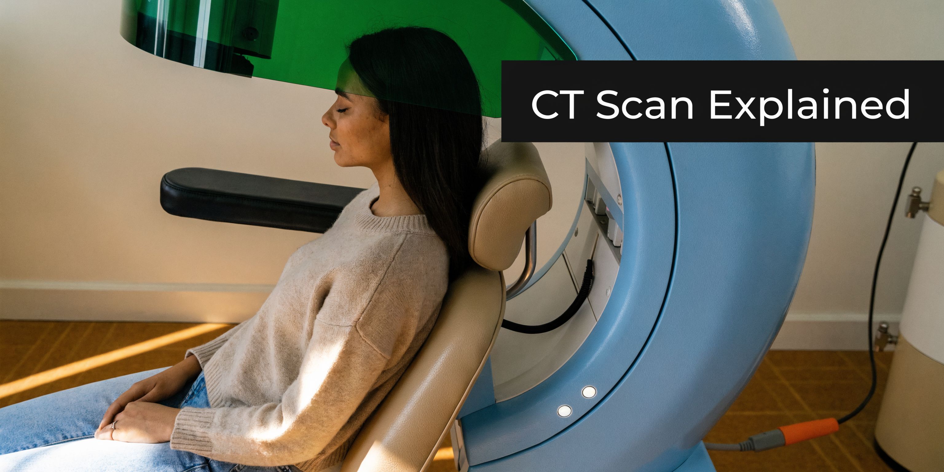
A CBCT scan works by capturing many images from different angles as the scanner rotates around your head. Software combines those images into a 3D view that your dentist can examine layer by layer.
The scan itself is quick. You stay awake, remain still for a short time, and the machine does the work around you. For many patients at our Piper Street clinic, the experience feels much easier than they expected once they see how open and straightforward the process is.
What appears on the screen is where the primary value begins. We can look at the area from the front, side, above, and in fine cross-sections, almost like checking the pages of a map book one by one instead of relying on a single snapshot.
What the scan can show
Because the image is three-dimensional, CBCT can reveal details that are difficult to judge on a flat view, such as:
- Bone height, width, and shape before implant planning
- Root position and root angle
- The course of important nerves in the jaw
- Impacted or unerupted teeth and how close they sit to other structures
- The relationship between upper teeth and the sinuses
- Areas that may suggest infection, damage, or unusual anatomy
- Airway shape in selected cases
For patients, that often means fewer surprises. If we can measure space properly before treatment starts, we can explain your options more clearly and plan with better precision.
How CBCT differs from a hospital CT
The word "CT" can sound intimidating, especially if you picture a large hospital scanner. Dental CBCT is built specifically for the mouth, jaws, and surrounding facial structures.
It focuses on a smaller area and is designed for dental planning rather than general medical imaging. That is one reason many Tamworth patients are relieved when they arrive and realise the scan is done standing or seated, with no tunnel and no needles.
Why dentists use it for planning
A good CBCT scan does more than create a sharp image. It gives measurements. That matters when a few millimetres can change a treatment decision, such as how close a tooth sits to a nerve or whether there is enough bone to support an implant securely.
If you would like to see how this fits into our wider approach to digital care, our page on advanced dental technology at Oasis Smiles Dental explains the tools we use to plan treatment with greater accuracy.
CBCT has become a well-established part of modern dental care. Analysts at Fortune Business Insights report that the global dental CBCT market was valued in recent years and is projected to keep growing through 2034, which reflects how widely this technology is now used in dental practices worldwide (Fortune Business Insights on the dental CBCT market).
For you, the practical takeaway is simple. A clearer view helps us explain your treatment in plain English, show you what we see, and make decisions with more confidence before any procedure begins.
The Clear Advantages Over Traditional Dental X-Rays
A common moment at our Piper Street clinic goes like this. A Tamworth patient points to a sore spot or a troublesome wisdom tooth and asks, "Can you see exactly what is going on?" Sometimes a standard X-ray answers that well. Sometimes it only shows part of the story.
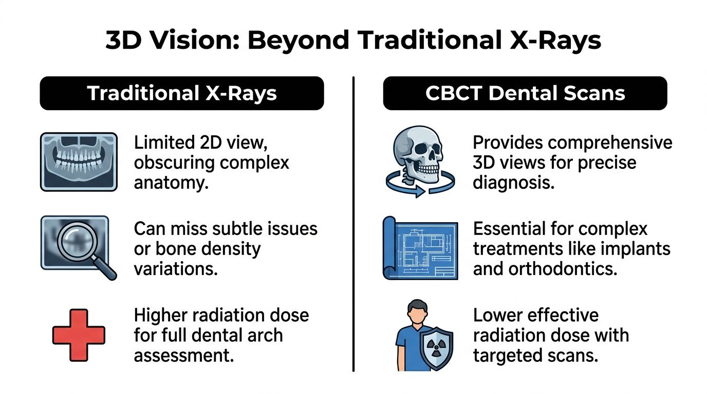
Traditional dental X-rays remain very useful for check-ups, simple decay, and many day-to-day concerns. But teeth do not sit in a flat row on a page. They sit in bone, beside nerves, under gums, and near the sinus. Once those structures overlap on a 2D image, judging depth can become much harder.
Flat picture compared with 3D map
A standard X-ray works a bit like a single photograph of a house. You can learn a lot from the front view, but you cannot easily tell what sits behind each wall.
A CBCT scan works more like a room-by-room map. It lets your dentist look through the area in thin slices and from different angles. That matters when a tooth is buried in the jaw, a root curves in an unusual direction, or an implant needs precise planning near a nerve. If you are exploring dental implant treatment options, that extra detail can make planning clearer and more predictable.
Side-by-side comparison
| Feature | Traditional dental X-rays | CBCT dental scan | |---|---|---| | View | 2D image | 3D image | | Best for | Routine checks and simple diagnosis | Complex planning and hidden anatomy | | Anatomy visibility | Can have overlap and distortion | Lets the dentist inspect structures from multiple angles | | Usefulness for surgery | Limited for depth and spatial planning | Strong for implants, wisdom teeth, and complex cases | | Airway review | Limited | Can support airway assessment in selected cases |
What this means for patients
For patients, a key advantage is clearer decision-making before treatment begins.
If a crack hides in a direction that a flat image does not show well, 3D imaging may reveal more. If a wisdom tooth sits close to a nerve, the relationship is easier to judge in three dimensions. If root positions matter for orthodontic planning, a CBCT scan can show details that a standard image may miss.
That extra visibility often leads to a simpler conversation in the chair. We can show you what we are seeing, explain why a treatment is recommended, and reduce the guesswork that can make dental decisions feel stressful.
What about radiation
This is a fair question, and many Tamworth patients ask it early.
A CBCT scan uses more radiation than a small routine dental X-ray, so we only recommend it when the added information is likely to help with diagnosis, planning, or safety. It still involves less radiation than a medical CT scan, which is one reason dental CBCT is useful for focused dental assessment rather than broad hospital-style imaging, as noted earlier in the article.
The goal is not to order more imaging. The goal is to choose the scan that fits the clinical question.
Why the right scan size matters
Scan size also matters. Dentists call this the field of view. In plain English, it means imaging the area we need, not a larger area that adds little value.
That approach follows the ALARA principle, which means keeping radiation as low as reasonably achievable while still getting the information needed for your care.
At Oasis Smiles Dental, the practical benefit is simple. If a regular X-ray can answer the question, we use that. If a 3D scan is the safer or clearer option, we explain why in plain language so you know what the scan adds and why it may be worth it.
How We Use 3D Imaging to Perfect Your Treatment
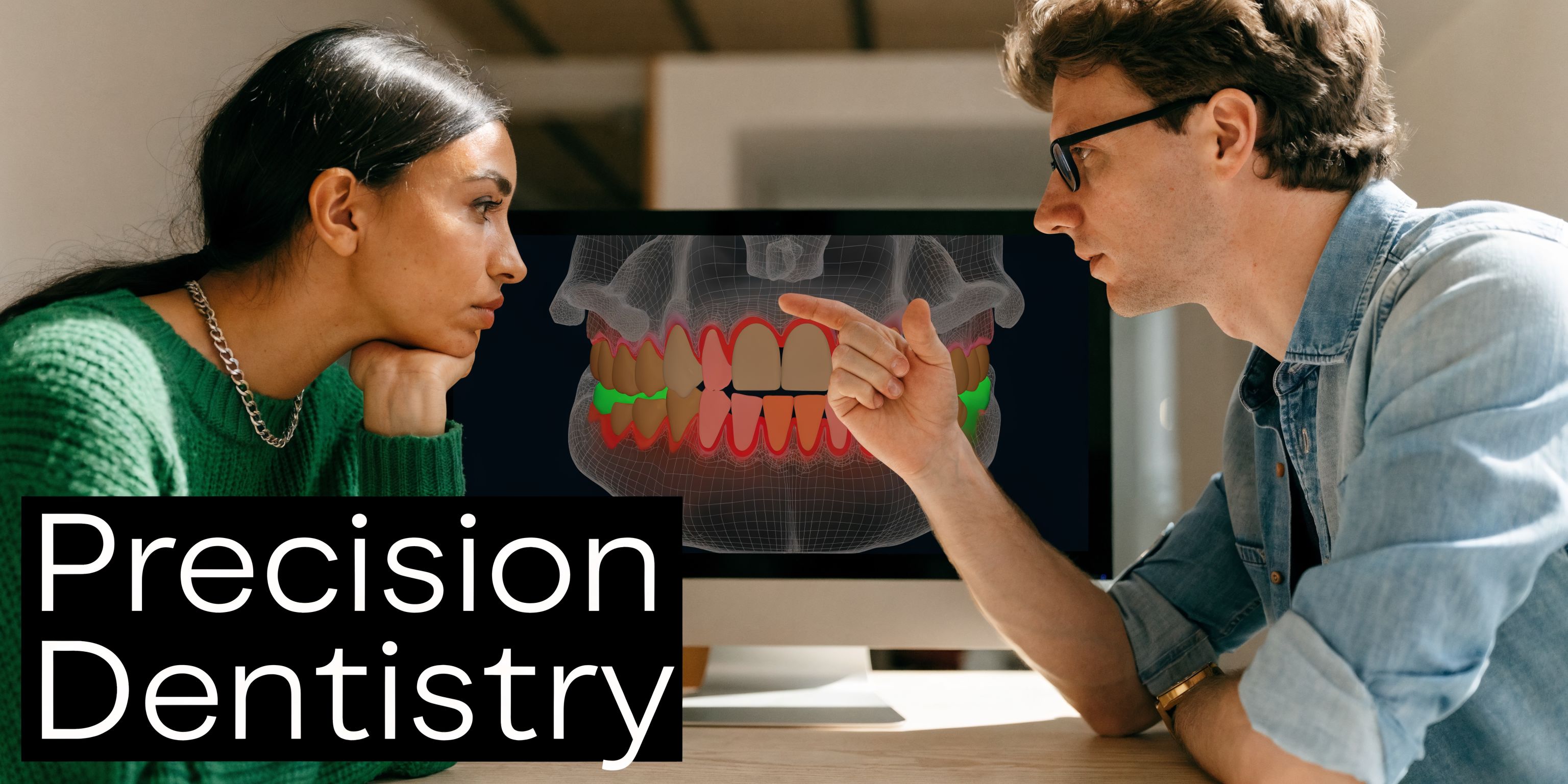
A 3D scan is most helpful when it changes the plan in a meaningful, practical way for you. At Oasis Smiles Dental in Tamworth, we use it like a detailed street map before a tricky drive. The more clearly we can see the roads, the easier it is to choose the safest route and explain it in plain language.
Dental implants with fewer unknowns
Implant planning is one of the clearest examples. A missing tooth can look straightforward from above the gum, but essential planning happens in the bone underneath. We need to check bone width, bone height, and the position of nearby structures such as the sinus or nerve canal.
A CBCT scan shows that area in three dimensions, which helps us plan the implant position, angle, and depth with much more confidence. For patients, that usually means a clearer discussion before treatment and fewer unwelcome surprises on the day. If you are exploring dental implant treatment options, our page on dental implants explains the restorative side of treatment in more detail.
Orthodontic planning that looks below the surface
Straight teeth are only part of the story. Roots matter just as much.
Teeth move through bone, and each root needs enough space and support. A 3D scan can help show impacted teeth, root positions, and areas where teeth are developing in awkward directions. That extra detail can be useful in complex orthodontic or clear aligner cases, especially when crowding makes a flat image less helpful.
For a patient, the benefit is easier to grasp than the technology. We can show you why a tooth may need a different approach, instead of asking you to trust a plan you cannot picture.
Root canal assessment when the cause is hard to spot
Some painful teeth are surprisingly hard to read on a standard X-ray. The symptoms are present, but the reason may stay hidden if the problem sits behind overlapping structures.
In selected cases, CBCT can reveal details such as:
- Missed canals that are difficult to see on a 2D image
- Root fractures that may change whether the tooth can be saved
- Infection around the root tip
- Unusual root shapes that affect treatment planning
We do not need a 3D scan for every root canal. We may recommend one when a case has failed before, symptoms do not match the usual picture, or the anatomy appears more complex than expected.
Wisdom teeth and surgical planning
Wisdom teeth often look simple on paper and less simple in practice. A standard X-ray can show that a tooth is trapped or angled. A CBCT scan can show how close it sits to the nerve canal, how it is tipped, and how much bone surrounds it.
That detail helps us discuss risk more clearly with Tamworth patients before a procedure. If the tooth is sitting close to a nerve, that matters. If it has a path that makes removal simpler than it first appeared, that matters too.
Airway assessment in selected cases
Some patients are surprised to hear that 3D dental imaging can also play a role in airway assessment. In selected cases, a CBCT scan can help show the shape and size of the airway more clearly than a flat image, which may support planning for an oral appliance or guide whether referral is appropriate.
The exact role of CBCT depends on the clinical question, and it does not diagnose sleep apnoea on its own. Sleep concerns still need proper medical assessment. We keep this part of care grounded and careful, especially if you have been told you snore, wake tired, or may be clenching and grinding at night.
Hidden findings that can change the plan
A scan is sometimes ordered because something does not quite add up. You may have swelling, delayed eruption, a tooth that behaves strangely, or a shadow on another image that needs a closer look.
In those situations, CBCT can help identify cyst-like areas, bone defects, unusual tooth paths, or other hidden findings that may change the next step. That is the primary value of 3D imaging at our Piper Street clinic. It helps us plan based on what is present, not on guesswork.
> When we can see the full shape of the problem, we can explain your options more clearly and plan treatment with greater confidence.
Your CBCT Scan at Oasis Smiles Dental What to Expect
For most patients, the actual scan is the easiest part. The worry beforehand is usually much bigger than the experience itself.
Here is what typically happens at a CBCT appointment at a modern dental clinic on Piper Street.
Before the scan
Preparation is minimal. You may be asked to remove items around the head and neck area, such as:
- Glasses if they are in the scan zone
- Earrings or necklaces
- Removable dental appliances
- Hair accessories if they contain metal
This is to reduce interference in the image.
If your visit relates to a surgical issue such as a buried or painful back tooth, a page like this one on wisdom teeth can help you understand why 3D imaging is sometimes part of the planning.
During the scan
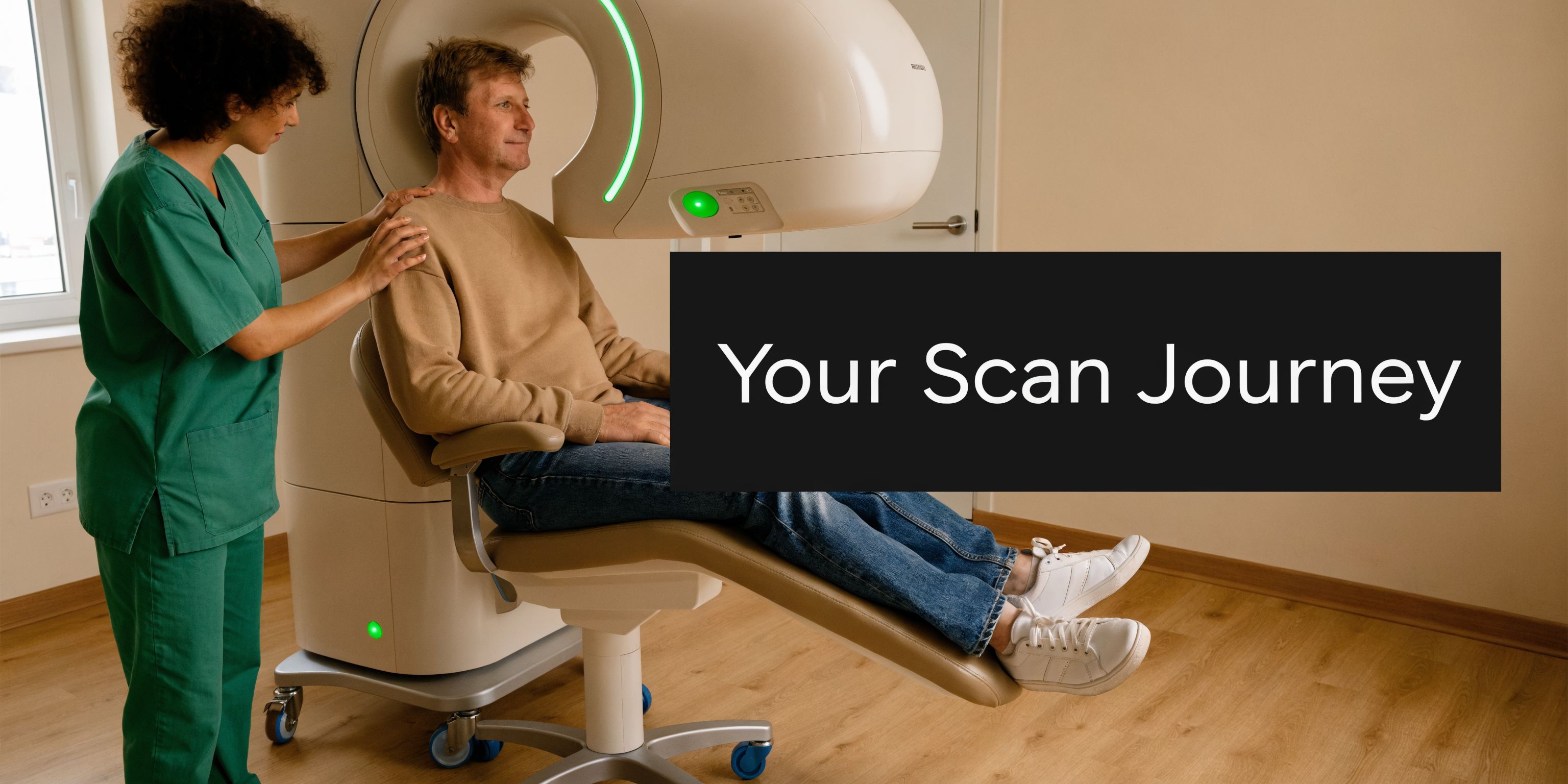
You will usually stand or sit in the machine, depending on the unit. A team member positions your head carefully, because stillness helps produce a clear image.
Then the scanner rotates around you. It does not touch you, and it does not feel enclosed like a hospital tunnel scanner.
The process is quick. Dental CBCT systems commonly capture many images in a short time in dental use, which is why patients often say, “That was it?” after the scan.
What the machine is doing
Although the scan feels simple, the technology is very refined. Technical reviews describe the X-ray tube as having a 0.5 mm focal spot, helping produce sharp images with voxels as small as 80 µm and resolution up to 5 line pairs per mm (technical review of CBCT image quality).
You do not need to remember those figures. The practical takeaway is that the machine is designed to pick up very fine detail in a small dental area.
After the scan
There is no recovery time. You can return to work, school, or your normal day straight after.
The images are then reviewed along with your examination findings and symptoms. In many cases, your dentist can show you the scan on screen and explain what they are seeing.
That conversation is often one of the most valuable parts. Instead of hearing a vague description, you can often see the position of the tooth, bone, root, or airway issue yourself.
> If you feel anxious before imaging, tell the team. A calm explanation and step-by-step guidance often makes the whole process feel much easier.
Costs Insurance and Accessibility in Tamworth
After “Will it hurt?”, the next question is usually, “What will it cost?”
That is a fair question, especially in regional areas where access to advanced imaging can feel less straightforward than in a major city.
Typical cost in regional NSW
In regional areas like Tamworth, Australian Medicare does not routinely cover dental CBCT scans, and out-of-pocket costs average AUD 200 to 400 per scan (regional CBCT access and cost discussion).
The exact fee can depend on the purpose of the scan and the size of the area being imaged. A focused scan for one site may differ from a broader treatment-planning scan.
Medicare, DVA, and health funds
Patients can get confused because “dental imaging” and “medical imaging” do not always follow the same rebate rules.
A few important points help:
- Medicare does not routinely pay for most dental CBCT imaging.
- DVA support may apply in eligible situations for veterans.
- Private health funds vary. Cover can depend on your extras policy and how the scan relates to treatment.
- Item rules matter. Eligibility often depends on the reason for the scan, not just the fact that a scan was taken.
That is why it is worth checking your specific cover before the visit. It can also help to review practical information about appointments and payment on the clinic’s patient information page.
Why local access matters
For people in Tamworth and the wider New England area, having advanced dental imaging available locally can make the process much easier. It can reduce extra travel, shorten the path between diagnosis and treatment planning, and make it simpler to discuss the results with the dentist who is providing your care.
That local convenience matters even more for families, older patients, and people who feel nervous about dental visits. Fewer moving parts often means less stress.
A practical way to think about value
A CBCT scan is not routine for every appointment, and it should not be. But when it changes the quality of diagnosis or planning, it can prevent uncertainty.
In practical terms, patients often value a 3D scan because it can help answer questions earlier, rather than discovering a problem halfway through treatment.
Frequently Asked Questions About Dental CT Scans
Is a dental CT scan dangerous
No. A dental CBCT scan is considered a safe diagnostic tool when it is clinically justified. Dentists do not order it for every visit. They use it when the extra detail is likely to improve planning or safety.
Will I need one every time I come in
No. Most routine appointments do not need CBCT imaging. Standard examinations and regular X-rays are often enough for check-ups, cleans, and straightforward treatment.
Does the scan hurt
Not at all. There are no needles, no drilling, and no physical contact from the scanner. You stay still while the unit rotates around your head.
Can fillings, crowns, or implants affect the image
Sometimes metal can create artefacts, which are areas of interference on the image. Dentists know how to interpret scans with that in mind, and they only request the scan when the likely benefit outweighs any limitation.
What if I already have old X-rays from another dentist
Old X-rays are often still helpful, but they may not answer the same question. A new CBCT scan may be needed if your dentist requires a 3D view of a specific area, updated anatomy, or more precise planning for current treatment.
Can I have a CBCT scan if I’m nervous
Yes. In fact, many nervous patients find the scan easier than they expected because it is quick and non-invasive. Let the team know if you feel anxious, and they can talk you through it step by step.
Your Next Step to Clearer Dental Care in Tamworth
Computed tomography dental imaging can sound technical, but the patient benefit is straightforward. It helps dentists see more clearly before they act.
That matters when treatment needs to be accurate, whether the issue involves an implant site, a wisdom tooth, a difficult root canal problem, orthodontic planning, or an airway assessment. A good CBCT scan can turn uncertainty into a clear plan.
For patients in Tamworth, that means better information, fewer surprises, and a calmer decision-making process. You are not being asked to trust guesswork. You are being shown a fuller picture of what is happening.
If you have been told you may need a 3D dental scan, the best next step is a conversation. Ask why it is being recommended, what it is expected to show, and how it will change your treatment plan. A clear answer should leave you feeling informed, not pressured.
---
If you’d like clear advice about 3D dental imaging, implants, wisdom teeth, sleep dentistry, or general family care, the team at Oasis Smiles Dental can help you understand your options in plain English and book a consultation in Tamworth.